Click here to view as a pdf: Calfhood Pneumonia When Is It Related To Ventilation

Dr. Ryan Leiterman holds degrees in both Agricultural Engineering and Veterinary Medicine
By Ryan Leiterman, D.V.M
Calfhood pneumonia can be frustrating to deal with. Understanding where the problem originates is the first step in creating a plan to combat it. Most cases of calfhood pneumonia can be placed into one of two broad categories: environmental causes vs. contagious causes.
Another way to look at these categories would be pneumonia cases caused by poor air quality vs. pneumonia cases caused by something other than air quality/ventilation.
Calfhood pneumonia is a complex, multifactorial disease that is rarely ever attributed to one factor. In the same way that spokes help a wheel keep its round shape when under a stress load, calves have six main “spokes” that help keep them healthy when subjected to stress. Those spokes are:
- Colostrum

- Calories
- Bedding
- Air Quality/ Ventilation
- Vaccination
- Sanitation
Each of the six “spokes” listed in Figure 1 are intertwined in a complex manner that helps keep calves healthy. Although ventilation and air quality are commonly implicated when discussing calfhood pneumonia, it is important to remember there are other factors that impact a calf’s respiratory health.
Environmental Pneumonia Cases ARE Typically Related To Air Quality And Ventilation
Poor ventilation in calf housing is the main environmental risk factor predisposing calves to respiratory disease.1 Cases of environmental pneumonia are by definition related to poor air quality and inadequate ventilation. Conventional wisdom would suggest that calves breathing in poor quality air would be inhaling large amounts of bacterial pathogens like Pasteurella multocida, Mannheimia haemolytica and Histophilus somni; and it’s the inhalation of these pathogens that cause disease. While this line of thinking logically makes sense, it is actually incorrect. The bacterial causes in most cases of environmental pneumonia do not actually come from the environment at all, they come from the calf itself.
Did you know that most healthy calves have Pasteurella, Mannheimia and Histophilus living in the upper part of their respiratory tract? According to Bradford Smith, “Mannheimia haemolytica, Pasteurella multocida and Histophilus somni are normal inhabitants of the nasal pharyngeal mucosa, but not the lung, and are considered “opportunistic pathogens.”2 Healthy calves carry these pneumonia-causing pathogens around in their nasopharynx (back of their throat) every day. While these bacteria may attempt to migrate down into the lung tissue, a healthy lung lining and strong immune system will keep these invaders at bay.
Poor air quality is defined as air that is high in contaminants such as noxious gasses (ammonia), particulates (dust), humidity and microorganisms. For calf barns, target ammonia levels less than 10 ppm and humidity levels between 50% and 80%.2
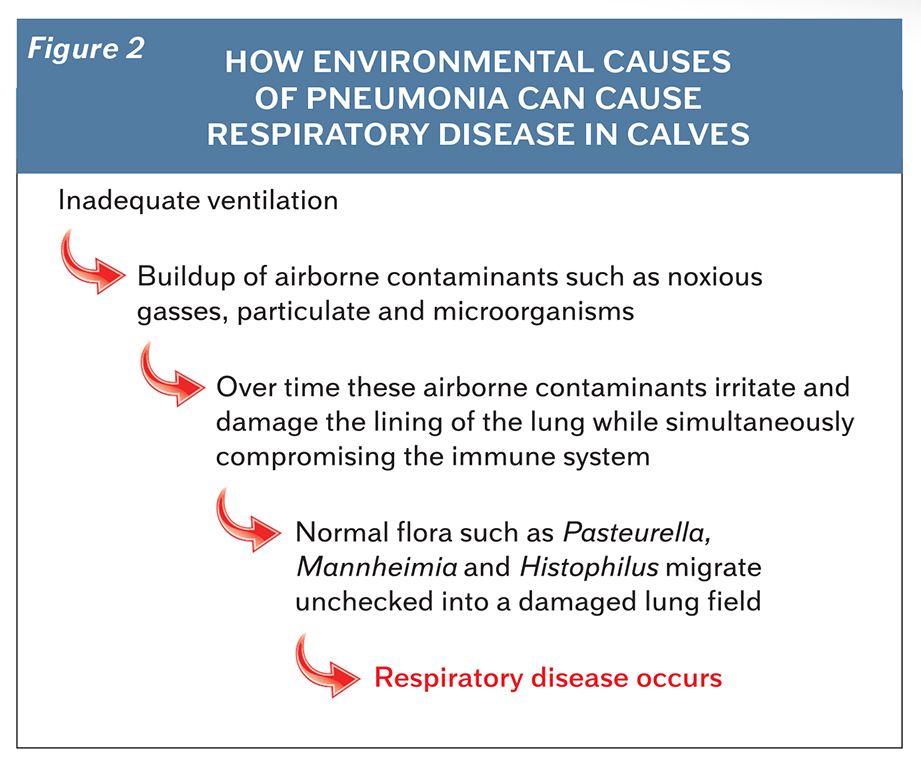
When these airborne contaminants are inhaled by calves in excessive levels, they act as an irritant to the lining of the respiratory system. Over time, exposure to these airborne irritants begins to damage the lining of the respiratory tract while simultaneously burdening the immune system. In the presence of poor air quality, normal flora like Pasteurella, Mannheimia and Histophilus can migrate down from the nasopharynx into the lung field that now has a compromised defense system, enabling pathogens to set up an infection and cause respiratory disease.
Calves housed in barns with individual pens can have a higher percentage of environmental pneumonia cases because many popular commercially available individual pens have solid plastic sides with mostly solid fronts and backs. While this style of penning reduces calf to calf contact which helps prevent contagious pathogen spread, it can restrict airflow inside the calf pen itself.
Environmental pneumonia cases can occur as a result of poor or inadequate ventilation and usually follow the pattern outlined in Figure 2.
If your farm is routinely dealing with calfhood pneumonia cases caused by Pasteurella, Mannheimia or Histophilus, revisit the 6 major spokes that make up the calf wheel of health paying particular attention to the air quality and ventilation system evaluation. A simple fogging test can tell you a lot about airflow throughout the barn (Figure 3).
Introducing fog next to the intake of an outside fan will demonstrate the path outside air takes once it enters the barn. Producers can also introduce fog in the center of a barn to see where it exhausts and how quickly it disperses. To estimate your barn’s air exchange rate, fill the barn with smoke and then time how long it takes for the smoke to disperse. Then take 60 divided by the time it takes for the smoke to clear (in minutes) and that will give you the number or air changes in an hour. For example, a barn that takes 10 minutes to clear the smoke would be 60 ÷ 10 = 6 air exchanges per hour. This smoke test can also help identify areas of still air, known as dead spots.

Contagious pneumonia cases are NOT typically related to air quality and ventilation. Pathogens such as Salmonella, Mycoplasma and Bovine Respiratory Syncytial Virus (BRSV) are not considered normal flora in healthy calves and are generally not associated with an airborne route of infection. These pathogens are found in infected animals and can be spread to uninfected animals through a variety of routes. The most common infection routes include exposure to an infected animal, infected colostrum or milk, contaminated feeding equipment and shared watering systems. Once a contagious cause of pneumonia, like Salmonella or Mycoplasma, is introduced into a facility it can be difficult to eradicate because the existing population of calves acts as a safe harbor for the pathogen; creating a situation where infected animals can pass the disease along to uninfected incoming animals. Group housed calves that use a continuous flow management style are at a higher risk of contagious disease transmission when compared to the “all-in/all-out” management method.
In 2011, Fiona Maunsell stated, “Once established in a multiage facility, Mycoplasma bovis is very difficult to eradicate, suggesting ongoing transmission from older to incoming calves…Transmission of Mycoplasma bovis in respiratory secretions is considered important in the epidemiology of infection.”3
Calves housed in barns with comingled group penning can have a higher percentage of contagious pneumonia cases. This is most likely explained because of the significant contact calves have with each other. While this style of housing generally allows for a more open pen style that promotes easier ventilation and improved air quality, things like nose to nose contact, shared feeding equipment and group waterers facilitate the spread of contagious pathogens throughout the group.
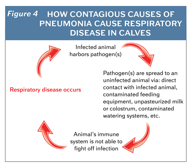 Contagious pneumonia cases occur as a result of exposure to infected animals or fomites transmitting infectious pathogens and follow the pattern outlined in Figure 4.
If your farm is routinely dealing with calfhood pneumonia cases caused by Salmonella, Mycoplasma and Bovine Respiratory Syncytial Virus (BRSV), revisit the 6 major spokes that make up the calf wheel of health with particular attention to sanitation protocols, colostrum management and vaccination evaluation.
There are countless sanitation products and protocols when it comes to keeping calf equipment clean. Regardless of the approach used, an effective cleaning and disinfection protocol should reduce pathogen buildup and remove biofilm from calf feeding equipment and penning. Execute the established cleaning and disinfection protocol, then use an ATP meter or surface protein swab to test what is being left behind. ATP meter readings of 200 RLU or less are the goal for calf feeding equipment and penning after they have been cleaned and disinfected.
Validate proper colostrum collection and storage by periodically performing colostrum cultures to evaluate bacteria levels. Consider additional culturing for Salmonella and Mycoplasma if there is a history of issues with these pathogens on your dairy. Confirm calves are utilizing the colostrum given and receiving the protection they need from it by routinely testing blood serum total proteins. Colostrum management practices are considered successful if 80 percent of calves tested are at or over a total protein level of 5.5 grams per deciliter.
Vaccine recommendations can vary due to regional and operational differences. There is no such thing as a one-size-fits-all vaccination protocol; therefore, it is best to consult with your veterinarian when designing a protocol for your operation. A judicious, effective vaccination protocol will limit vaccine use to those vaccines with proven efficacy.
Remember that few things in life are black and white. Calfhood pneumonia cases are often complex with multiple confounding factors. Addressing a calfhood respiratory disease problem is more complex than just saying “We need better ventilation in this barn.” Investing in a better ventilation system will pay back dividends when struggling with an environmental pneumonia problem, but will do little to reduce respiratory disease rates when the cause is contagious in nature. Before spending money on changing the ventilation system, be sure that poor air quality is really the issue at hand.
This article was originally published with the Progressive Dairy Magazine at https://www.progressivedairy.com/topics/calves-heifers/calfhood-pneumonia-when-is-it-related-to-ventilation-and-when-is-it-something-different